
Medical Tourism Blog
Ptosis Correction in Korea: Methods, Recovery, Risks and Travel Planning
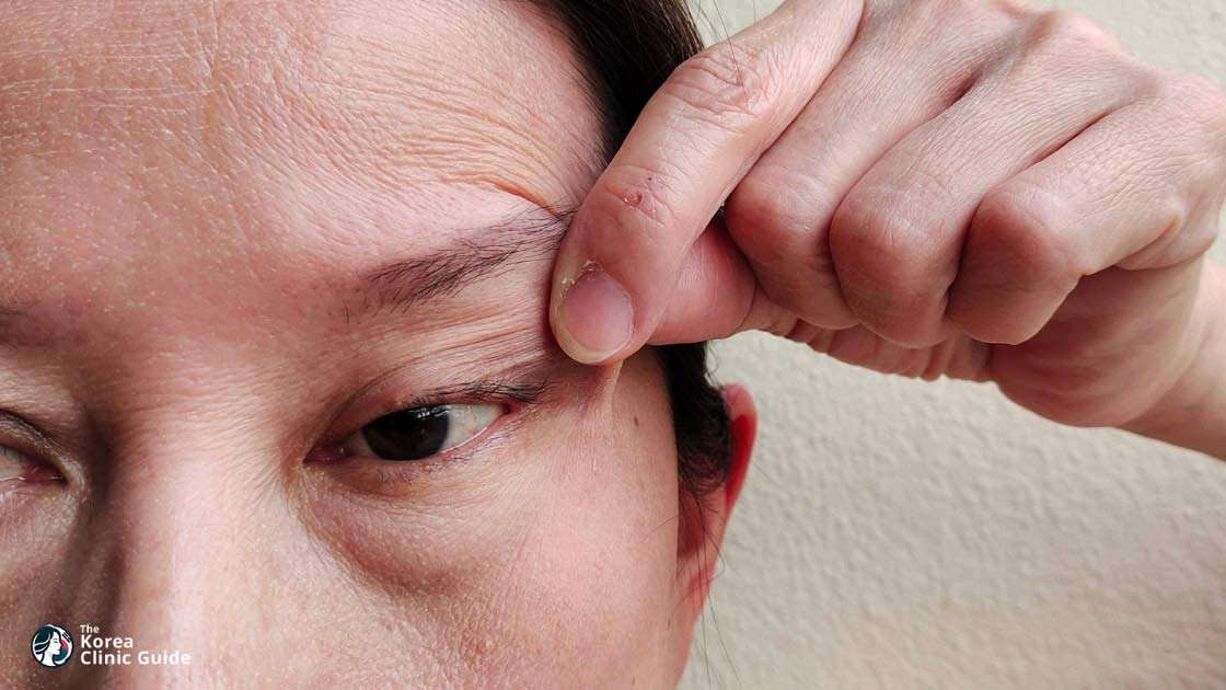
Table of contents
- Ptosis Correction at a Glance
- What Is Ptosis Correction?
- Who Is a Good Candidate for Ptosis Correction?
- How Is Ptosis Correction Performed?
- Why Visit Korea for Ptosis Correction?
- Key Information for International Patients
- Which Are the Best Clinics in Korea for Ptosis Correction?
- How Much Does Ptosis Correction Cost in Korea?
- Ptosis Correction From Start to Finish
- Alternatives to Ptosis Correction
- How Can I Prepare for Ptosis Correction?
- Aftercare Advice
Considering treatment in Korea? Everything you need to know e.g. — how to avoid scams, visas, interpreters, recovery tips — in our Medical Tourism Master Guide. Plan with confidence in minutes, not weeks!
Last updated: September 2026
Ptosis Correction at a Glance
- Purpose — Ptosis correction is an outpatient eyelid operation that raises an abnormally low upper-lid margin to improve vision obstruction, brow strain, chin-up posture, or persistent asymmetry.
- Cost in Korea — Typical pricing is ₩1,500,000–₩4,500,000 (about US$1,100–$3,300), with the final quote affected by technique, one versus two eyelids, anesthesia, and whether a frontalis sling or thigh-fascia harvest is needed.
- Technique is individualized — External levator repair is common for age-related ptosis with good levator function; internal MMCR suits selected mild-to-moderate cases, while severe poor-function ptosis may require a frontalis sling using brow-muscle lift.
- Recovery and results — Swelling, bruising, tearing, light sensitivity, and ointment-related blur are usually most noticeable for 1–2 weeks; lid height and contour continue settling for weeks to months, so final symmetry should not be judged early.
- Travel planning — International patients should generally plan 8–10 days in Korea for an uncomplicated adult repair, including a review and possible suture removal at about 1 week; fascia-lata sling cases or substantial swelling may require about 14 days.
- Key safety limitation — A persistently open eyelid can cause dry eye and corneal exposure, while undercorrection, overcorrection, asymmetry, recurrence, and revision are possible; new, sudden, or fluctuating droop needs medical evaluation before elective surgery.
- Korea-specific consideration — Korean ophthalmic practice documents access to levator repair, levator resection, and frontalis suspension, and registered foreign-patient institutions must carry malpractice liability insurance; neither point proves superior outcomes versus other countries.
What Is Ptosis Correction?
Ptosis correction is eyelid surgery that raises an abnormally low upper eyelid margin. It aims to clear vision obstruction, reduce brow strain or chin-up posture, and improve eyelid symmetry while maintaining safe eye closure. It is a surgical procedure, usually performed as an outpatient operation.
Who Is a Good Candidate for Ptosis Correction?
- Stable functional droop — Adults with a consistently low upper-lid margin that blocks vision, causes brow strain or a compensatory chin-up posture, or creates persistent eyelid asymmetry after proper assessment.
- Acquired aponeurotic ptosis — People with age-related stretching or disinsertion of the levator aponeurosis, often with good levator function, are commonly assessed for levator repair.
- Congenital ptosis — Children need pediatric ophthalmology or oculoplastics assessment when the lid blocks the visual axis, creates a sustained chin-up posture, or risks amblyopia (lazy-eye development). Congenital ptosis can threaten visual development.
- Realistic surgical goals — Good candidates understand that the priority is a safely elevated lid with corneal protection and improved symmetry, not perfectly identical eyelid height or crease shape.
New, sudden, or fluctuating ptosis requires medical evaluation before elective surgery, especially with double vision, unequal pupils, headache, neck pain, weakness, abnormal eye movements, or breathing or swallowing symptoms; these can indicate urgent neurologic disease. Third-nerve palsy, Horner syndrome, and myasthenia gravis require diagnosis rather than cosmetic treatment. Delay repair for active eye infection, uncontrolled dry eye, poor tear production, weak Bell phenomenon, or inadequate corneal protection, because raising the lid can worsen exposure keratopathy. Thyroid eye disease, progressive muscle disease, unstable myasthenia gravis, and poorly controlled systemic illness need stabilization first. A full examination must also distinguish true ptosis from pseudoptosis caused by excess upper-lid skin, brow descent, or facial asymmetry.
How Is Ptosis Correction Performed?

Technique selection follows the cause and degree of droop, levator function, MRD1 measurement, brow position, ocular-surface health, previous surgery, and whether one or both lids are involved. Incision preference alone is not a sound basis for choosing a technique.
- External levator advancement — Through an upper-lid-crease incision, the surgeon reattaches or advances a stretched levator aponeurosis. This is a common approach for acquired aponeurotic ptosis with good levator function and permits intraoperative adjustment of lid height and contour.
- Levator resection — The levator muscle is shortened or strengthened when it still functions but is weaker than needed. It is often used for congenital ptosis with fair levator function.
- MMCR posterior repair — Müller muscle-conjunctival resection (MMCR) is performed from inside the eyelid for selected mild-to-moderate cases with good levator function, often after phenylephrine testing. A meta-analysis of seven comparative studies involving 1,038 eyelids found similar postoperative MRD1 and undercorrection rates to external levator advancement, with lower overcorrection and reoperation rates for MMCR; this does not make it suitable for every form of ptosis.
- Frontalis sling or frontalis advancement — Used when levator function is very poor, particularly in severe congenital or myogenic ptosis. The brow muscle provides the lifting force, using autologous thigh fascia lata or synthetic sling material. Incomplete blink, lagophthalmos, corneal exposure, recurrence, and revision are important tradeoffs.
For congenital ptosis, a 2023 review of 14 randomized trials covering 909 eyes found that procedures and sling materials affect different outcomes, including lid height, lagophthalmos, and cosmetic symmetry. Evidence remains heterogeneous, so severe congenital, neurogenic, myogenic, revision, and combined aesthetic cases require individualized planning.
Why Visit Korea for Ptosis Correction?
Korean ophthalmic practice documents access to external levator repair, levator resection, and frontalis suspension rather than a single standard operation. This matters because true ptosis treatment depends on the lifting mechanism and corneal-protection risk. Current evidence does not establish that Korea produces superior ptosis outcomes compared with other countries.
- Documented surgical spectrum — A 24-year Seoul tertiary-referral cohort reported use of aponeurosis repair, levator resection, and frontalis suspension across acquired and congenital cases, supporting the value of selecting a surgeon who can assess and treat different mechanisms.
- Mechanism-focused assessment — In that cohort, acquired ptosis was commonly aponeurotic with good levator function, while congenital cases were more often moderate-to-severe with fair-to-poor function. These distinctions support measurement-led planning rather than treating every low lid as a cosmetic crease problem.
- Foreign-patient registration framework — Korea’s Ministry of Health and Welfare registration system requires registered foreign-patient institutions to carry malpractice liability insurance. The Medical Korea registration framework is an administrative and patient-safety safeguard, not proof of better surgical results; confirm registration, interpretation, coordination, and post-departure support directly with the facility.
- Evidence limitation — The Korean surgical cohort was retrospective, single-center, and covered historical cases from 1991–2014. It is not an international comparison and cannot predict an individual patient’s result or revision risk.
Key Information for International Patients
- Minimum stay — Plan at least 8–10 days for straightforward adult eyelid-only repair, allowing an early review and a suture check or removal at about 1 week. Plan about 14 days for a frontalis sling using thigh fascia lata, bilateral surgery with substantial swelling, complications, or surgeon-requested observation.
- Flight timing — There is no universal evidence-based flight interval specific to ptosis repair. Do not leave Korea until the first review confirms acceptable vision, wound status, corneal protection, and adequate eyelid closure.
- Recovery and itinerary — Bruising, swelling, soreness, tearing, light sensitivity, and ointment-related blurred vision are most noticeable during the first 1–2 weeks. Avoid demanding sightseeing, driving, and activities requiring clear vision during this period.
- Repeat visits and revision — Lid height, contour, and early asymmetry continue settling for weeks to months. Some patients need later adjustment or revision; in the cited Korean tertiary cohort, 3-year cumulative reoperation was 15.7% for congenital and 10.4% for acquired ptosis, figures that reflect that center’s historical case mix rather than an individual prediction.
- Remote follow-up — Obtain an English operative report stating diagnosis, laterality, technique, sling material where relevant, anesthesia, prescribed medicines, and an urgent-contact pathway. Arrange remote review before departure and identify a local ophthalmologist or oculoplastic surgeon at home.
Travel note: Schedule flexible flights and accommodation close to the clinic. A persistently open eye, significant dryness, new double vision, or a vision change needs prompt examination, not remote reassurance alone.
Which Are the Best Clinics in Korea for Ptosis Correction?
Listed below are some of the best clinics in Korea for ptosis correction.
1. THEPLUS Plastic Surgery
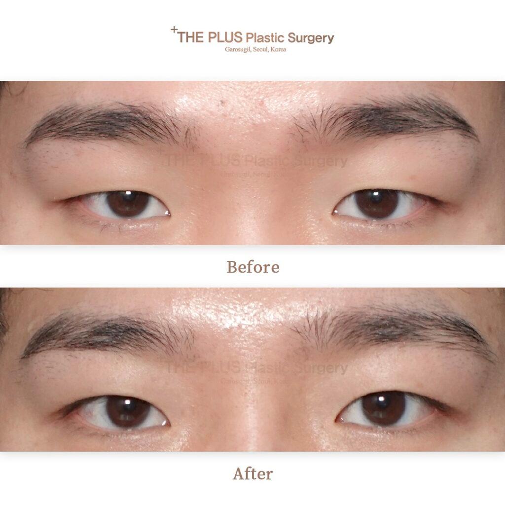
At THEPLUS Plastic Surgery, ptosis correction is offered to lift drooping upper eyelids by correcting the muscles that open your eyes, with the aim of reducing a heavy or tired appearance and creating a brighter-looking eye area.
- URL: THEPLUS Plastic Surgery Website
- Location: Gangnam-gu, Seoul
- Procedure focus:
- Ptosis correction addresses upper-eyelid drooping through muscle correction.
- Double-eyelid surgery and blepharoplasty are also available for eyelid-crease definition or adjustment of excess eyelid skin and tissue.
- Surgical approach:
- The clinic describes its aesthetic approach as seeking harmony with your existing facial features and natural-looking balance.
- Its surgeons include board-certified plastic surgeons and physicians active in Korean and international plastic-surgery societies.
2. Made Young Plastic Surgery
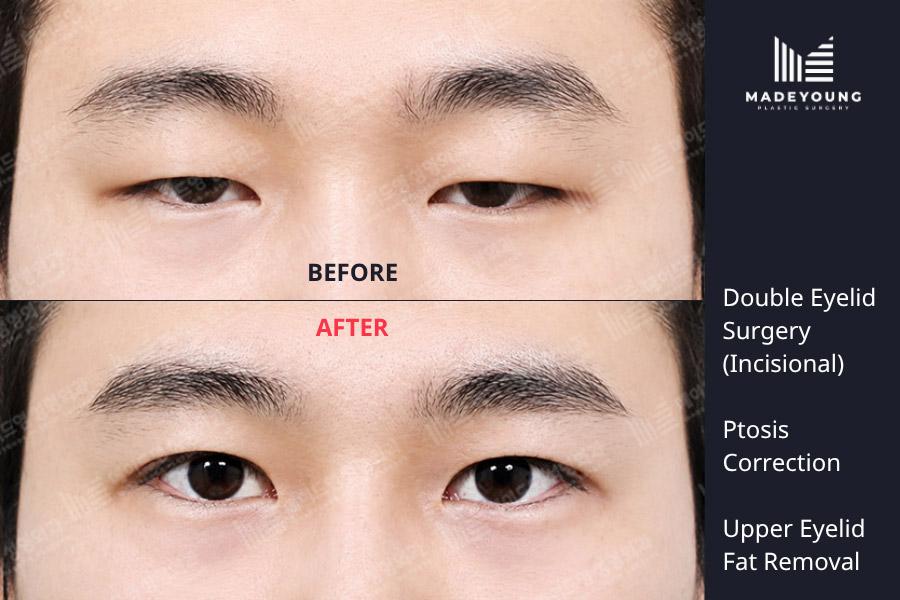
At Made Young Plastic Surgery, your ptosis correction can be planned around customized eyelid details, including incision lines, double-eyelid height, eyelid thickness, and your overall desired eye image. The procedure repairs drooping eyelids by tightening the muscle that lifts the eyelid.
- URL: Made Young Plastic Surgery Website
- Location: Seocho-gu, Seoul
- Eye-surgery experience:
- Dr. Park Byung-Chan is described as having 15 years of experience dedicated exclusively to eye surgery.
- You receive direct consultation and diagnosis from the medical team before treatment.
- Safety and recovery:
- Full-time board-certified anesthesiologists are on site, with one anesthesiologist assigned to continuous monitoring of one person during procedures.
- The clinic describes an emergency cross-check system involving multiple anesthesiologists and CCTV coverage of procedures.
- A separate aftercare center provides systematic post-procedure care.
- Related eye options: Available eye procedures include non-incisional and incisional double-eyelid surgery, upper and lower blepharoplasty, canthoplasty, and dark-circle treatment.
3. RINE Plastic Surgery Clinic
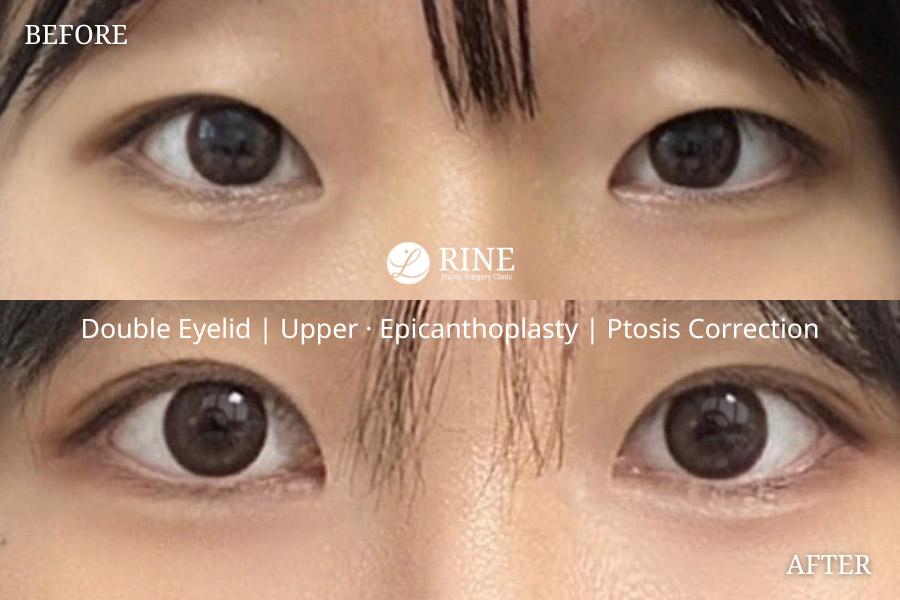
At RINE Plastic Surgery Clinic, ptosis correction strengthens the levator muscle to address drooping upper eyelids and improve iris exposure; it may also be combined with double-eyelid surgery. Your plan is designed individually around your facial proportions, skin condition, and eye-line goals.
- URL: RINE Plastic Surgery Clinic Website
- Location: Seocho-gu, Seoul
- Director-led care:
- Director Jang Nam personally handles consultation, surgery, treatment, and recovery rather than transferring these stages between providers.
- The clinic reports that its director has personally performed more than 10,000 eye surgeries and has over 15 years of clinical experience.
- Procedure planning:
- Ptosis correction is intended to improve both the functional and aesthetic appearance of drooping eyelids.
- You receive a 1:1 customized eye-line design intended to produce a natural, harmonious result.
- Non-incisional double-eyelid surgery, canthoplasty, and revision eye surgery are available when relevant to your eye shape or previous surgery.
- Facilities and aftercare:
- The clinic cites a Stryker full-HD endoscope, FDA-approved genuine Endotine, and WhiteLite bipolar equipment.
- You can recover in a private single-person recovery room.
- The clinic describes continuous postoperative care, infection-control measures, surgical safety protocols, and emergency preparedness.
How Much Does Ptosis Correction Cost in Korea?
Ptosis Correction is supplied at ₩1,500,000–₩4,500,000 (approximately US$1,100–$3,300). Individual quotes vary because the repair method, number of eyelids treated, anesthesia requirements, and need for additional tissue work differ by diagnosis and eyelid function.
| Procedure Price | Korean Won (₩) | USD ($) |
|---|---|---|
| Low Price | ₩1,500,000 | $1,100 |
| High Price | ₩4,500,000 | $3,300 |
Exchange rate as of 2026-09-06: 1 KRW = 0.000741 USD.
What Affects the Price?
- Repair technique and levator function — Selected mild-to-moderate cases may be suitable for posterior MMCR, while external levator advancement or levator resection requires direct work on the eyelid elevator through an incision. Severe ptosis with very poor levator function may require frontalis suspension or frontalis advancement, a more involved reconstruction that uses the brow muscle to lift the lid.
- Frontalis sling material and thigh harvest — A frontalis sling can use synthetic material or autologous fascia lata. Using fascia lata requires harvesting tissue from the thigh in addition to the eyelid repair, adding a second operative site, operative time, consumables, and wound care requirements.
- One eyelid versus bilateral correction — Treating both upper lids requires surgery across two eyelids and assessment of height, contour, blink, and symmetry on each side. Bilateral repair therefore uses more operating time and surgical resources than a straightforward unilateral repair.
- Anesthesia and patient complexity — Many adult eyelid-only repairs are performed with local anesthesia, sometimes with monitored sedation. Children and some complex cases require general anesthesia, which adds anesthesia staffing, monitoring, recovery resources, and facility use.
- Revision or exposure-risk management — Revision ptosis repair, substantial pre-existing dry eye, weak eye closure, or corneal-exposure risk requires more careful intraoperative adjustment and postoperative monitoring to achieve lid elevation without compromising corneal protection.
Cost tip: Request an itemized quote stating what is included, particularly the planned technique, laterality, anesthesia, facility use, sling material or fascia-lata harvest when relevant, medications or consumables, and postoperative reviews or suture removal.
The US$1,100–$3,300 conversion is approximate, based on the supplied exchange rate dated 2026-09-06. Check the exchange rate again when budgeting.
Ptosis Correction From Start to Finish
- Remote screening and communication — Send straight-ahead photographs with relaxed brows, the onset and pattern of droop, previous eyelid or eye procedures, contact-lens use, dry-eye symptoms, medical diagnoses, medication list, and any visual-field testing. Confirm English consultation or interpretation, the expected stay, and follow-up arrangements. Photographs cannot establish the final surgical plan.
- In-person ophthalmic assessment — The surgeon checks visual acuity, pupils, eye movements, ocular surface, tear function, brow compensation, facial asymmetry, MRD1, lid-crease height, and levator excursion. Ptosis assessment distinguishes aponeurotic, mechanical, neurologic, and myogenic causes. Phenylephrine testing can help select a posterior approach in appropriate cases.
- Medical clearance and surgical planning — Findings of fluctuating ptosis, abnormal pupils, eye-movement limitation, or systemic muscle weakness prompt medical or neuro-ophthalmic investigation. The surgeon confirms whether the problem is true ptosis, pseudoptosis, or a combination with excess skin or brow descent.
- Anesthesia and repair — Adult surgery is usually outpatient under local anesthetic, sometimes with monitored sedation. Children and some complex cases require general anesthesia. The surgeon advances or shortens the levator, performs MMCR from inside the lid, or connects the lid to the frontalis muscle; lid height and contour are checked before closure.
- Discharge on the day of surgery — Use prescribed lubricant and antibiotic ointment or drops as directed. Ointment can blur vision. Arrange an adult escort after sedation or general anesthesia and do not drive yourself home.
- Early clinic review — Attend the surgeon’s review before international departure. Visible skin sutures are commonly removed at about 1 week, while some sutures are buried. The clinician checks lid closure, corneal surface, vision, swelling, and wound healing.
- Overseas follow-up — Send the clinic requested photographs after returning home and keep the operative report accessible. Seek local urgent ophthalmic assessment for vision loss, severe pain, marked exposure symptoms, infection concern, or rapidly worsening swelling.
Alternatives to Ptosis Correction
The correct alternative depends on whether the low appearance comes from the eyelid margin itself, excess skin, brow descent, or an underlying disease. Non-surgical options avoid an operation but do not restore levator function; skin- or brow-focused surgery can improve pseudoptosis but does not reliably correct true ptosis.
Observation and cause-directed care
- Stable mild ptosis — Observation is reasonable when there is no visual obstruction and the condition is stable.
- Underlying disease treatment — Myasthenia gravis, thyroid eye disease, neurologic disease, inflammation, or an eyelid mass requires disease-specific treatment before considering elective lid-position surgery.
Ptosis-crutch glasses
- Mechanical support — A bar attached to eyeglasses can hold the upper lid higher for selected people who cannot or do not wish to undergo surgery.
- Tradeoff — This does not correct the cause and can be inconvenient, particularly with dry eye or contact-lens use.
Upper blepharoplasty
- Excess-skin treatment — Upper blepharoplasty removes or repositions excess upper-lid skin when dermatochalasis creates a heavy or obstructed appearance.
- Key limitation — It does not repair a weak or detached eyelid elevator and can leave true ptosis untreated.
Brow or sub-brow lift
- Brow-descent treatment — An eyebrow lift or subbrow lift can address brow descent that pushes skin onto the upper lid.
- Key limitation — These procedures do not reliably elevate an abnormally low eyelid margin caused by true ptosis.
How Can I Prepare for Ptosis Correction?
- Disclose medical and eye history — Report the onset or fluctuation of ptosis, double vision, dry eye, glaucoma, thyroid eye disease, myasthenia gravis, diabetes, prior eye or eyelid surgery, contact-lens use, and planned LASIK or LASEK. Ptosis repair can alter corneal topography and refraction, so eyelid position should stabilize before refractive surgery.
- Review blood-thinning medicines — Tell the surgeon and prescribing clinician about aspirin, ibuprofen, naproxen, warfarin, apixaban, clopidogrel, and all other anticoagulant or antiplatelet drugs. Do not independently stop prescribed medicines; changes require approval from both clinicians.
- Pause supplements only when instructed — Ask whether to stop fish oil or omega-3, vitamin E, ginkgo, garlic, ginseng, turmeric or curcumin, flaxseed oil, and St John’s wort. Many perioperative protocols request a 1–2 week pause because of bleeding, bruising, or anesthesia-interaction concerns.
- Stop nicotine and limit alcohol — Stop smoking and vaping well before surgery and remain nicotine-free through healing because nicotine impairs wound healing. Avoid alcohol around surgery and while taking sedating medicines when instructed.
- Follow fasting instructions exactly — Use the anesthetist’s written fasting schedule when sedation or general anesthesia is planned; do not rely on a generic rule.
- Prepare practical supplies — Bring glasses rather than contact lenses, loose front-opening clothing, cold compresses, and surgeon-approved lubricating drops if advised. Arrange a companion for the first night after sedation, bilateral surgery, limited vision in the other eye, or pediatric care.
Travel note: Book flexible flights and stay close enough to attend the early review and possible suture removal. Avoid strenuous sightseeing during the first postoperative week.
Confirm the operating clinic’s medication, fasting, companion, accommodation, and travel requirements in writing; the treating doctor’s individualized instructions take precedence.
Aftercare Advice
Protect the ocular surface and attend the early review: a raised lid that does not close fully can expose and damage the cornea.
- Lubrication and prescribed medicine — Use antibiotic ointment, drops, and lubricating drops or ointment exactly as prescribed. Ointment commonly causes temporary blurred vision; lubrication is particularly important when blink or closure is incomplete.
- Swelling control — Apply cold compresses as instructed and sleep with the head elevated. Bruising and swelling commonly last 10–14 days, while stiffness, minor asymmetry, and residual swelling can take weeks to months to settle.
- Protect the eyelids — Do not rub the eyes or disturb sutures. Avoid dusty or dirty environments, swimming, eye makeup, and contact lenses until the surgeon clears them; guidance commonly restricts these for about 2 weeks, with contact lenses sometimes delayed up to 4 weeks when dryness persists.
- Limit pressure-raising activity — Avoid bending, heavy lifting, vigorous exercise, and strenuous activity for about 2 weeks unless the surgeon gives a different schedule. Do not drive until vision is clear and no sedating medication is affecting alertness.
- Know urgent warning signs — Seek emergency ophthalmic assessment for reduced or lost vision, escalating severe pain, rapidly increasing tense swelling or bleeding, fever, pus, spreading redness, new double vision, or a persistently open painful and light-sensitive eye. Serious orbital bleeding with vision loss is rare but time-critical.
- Judge results at the right time — Do not assess final lid height or contour in the first few weeks. Early asymmetry is common, and later adjustment is sometimes necessary.
Follow the treating doctor’s instructions over general guidance, and contact the clinic promptly when healing, vision, comfort, or eyelid closure appears abnormal.
Frequently Asked Questions
Related Blogs

Upper Blepharoplasty in Korea: Methods, Recovery, Risks, and Travel Planning
Upper blepharoplasty conservatively removes excess upper-lid skin and, when indicated, limited muscle or fat after assessment of ptosis, brow position, eyelid closure, and eye health. International patients should plan a 7–10-day stay in Korea, attend early follow-up, arrange flexible travel and aftercare, and seek urgent assessment for vision changes or severe swelling.
Updated: Oct 13, 2026
Lower Canthoplasty in Korea: Methods, Recovery, Risks, and Travel Planning
Lower canthoplasty includes lower-eyelid-lowering and lateral canthal techniques, with candidacy based on lid support, ocular-surface health, and realistic anatomical goals. International patients can find guidance on technique clarification, 8–10 day travel planning, follow-up, aftercare, and urgent eye symptoms.
Updated: Oct 13, 2026
Upper Canthoplasty in Korea: Methods, Recovery, Risks and Travel Planning
Upper canthoplasty releases or redrapes the upper-medial epicanthal fold to improve visibility of an upper-eyelid crease, often alongside double-eyelid surgery. The article covers candidacy, surgical approaches, recovery, aftercare, travel planning for Korea, and warning signs requiring urgent eye care.
Updated: Oct 13, 2026
Epicanthoplasty in Korea: Methods, Recovery, Risks and Travel Planning
Epicanthoplasty is an outpatient procedure that releases and redrapes inner-eye-corner tissue to permanently reduce an epicanthal fold, with technique selected for anatomy, scar risk, and treatment goals. International patients should plan a 7–10-day stay for assessment and early wound review, follow aftercare restrictions, and arrange local eye-care follow-up after travel.
Updated: Oct 13, 2026
Clinics offering Ptosis Correction
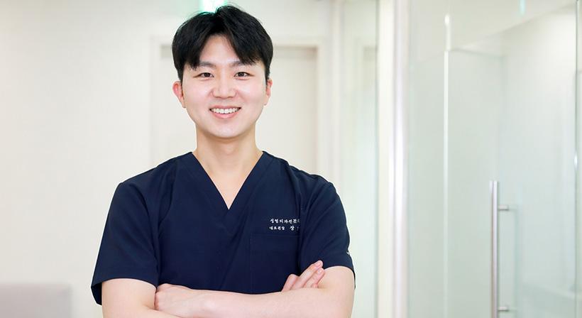
RINE Plastic Surgery Clinic | Facelift and Eye Rejuvenation Surgery
Nonhyeon

SH Clinic: Comprehensive Women's, Men's & Couples' Health Services in Korea
Sinsa

THEPLUS Plastic Surgery Korea | Eye, Nose, Face & Body Surgery
Sinsa

Made Young Plastic Surgery: Rejuvenating Aesthetic Excellence in Korea for Face Lifts, Contouring and More
Gangnam Station